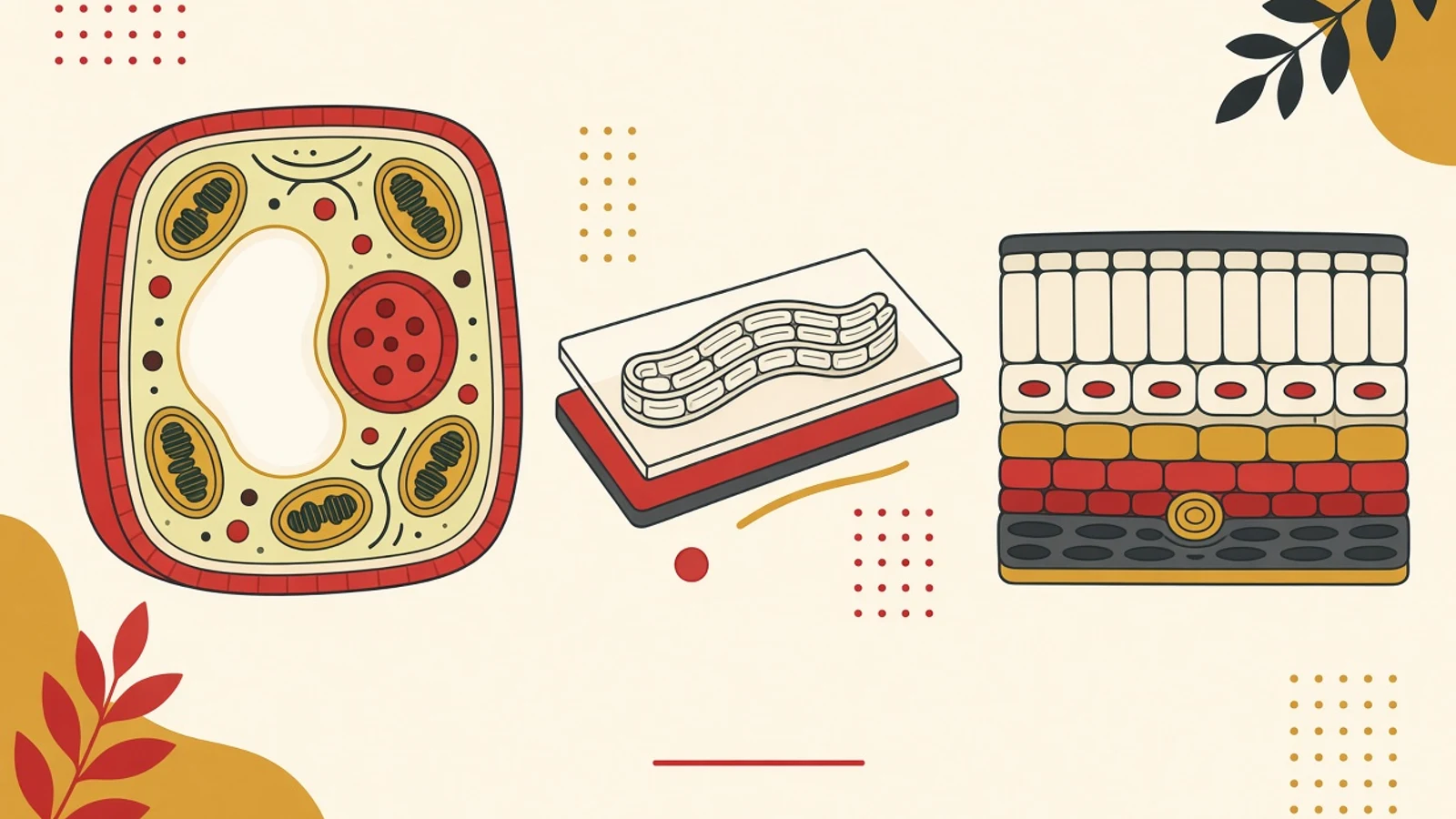
The least tiring way to study cells and tissues is to treat a cell as a small factory where every organelle holds one job: mitochondria produce ATP, ribosomes assemble proteins. Once that job map is solid, you move up to tissues and then test everything through three simple observations: a cheek cell, a thin layer of onion skin, and a Rhoeo leaf.
- Learn each organelle through its job, then sort it under one of three labels: shared by plant and animal cells, plant-only, or mostly animal-only.
- Plant tissues split into meristems that keep dividing and permanent tissues; animal tissues come down to four basic types: epithelial, connective, muscle, nervous.
- Cheek cells, onion epidermis and Rhoeo leaves move the topic off the textbook page and into images you have seen with your own eyes.
Three numbers that make cell scale feel real
Why cell organelles stick better when you picture a factory
The cell chapter in high school biology feels heavy because it arrives as a roll call of names: endoplasmic reticulum, Golgi apparatus, peroxisome, centrosome, and then the cell wall and central vacuole on top. A roll call like that evaporates soon after the test. What lasts in memory is a picture with a workflow, and a cell genuinely runs like a small factory. It has a gate where materials enter and leave, an archive holding the blueprints, an assembly line, a workshop, a packing department, a power plant, and a cleaning crew. The analogy is a memory aid, so every mapping has to match the function printed in your textbook. The plasma membrane works as the gate because its phospholipid bilayer and proteins separate the cell's contents from the outside while controlling what passes in and out. The nucleus works as the archive because it stores DNA and directs protein production. If you ever find a mapping that clashes with the real function, drop the analogy and keep the textbook function. Scale belongs in your head from the start too. Eukaryotic cells usually measure 10 to 100 micrometres, while prokaryotic cells measure 0.1 to 5 micrometres (OpenStax Biology 2e, §4.2). There is a reason they stay small: as a cell grows, its surface area to volume ratio shrinks. A cube with 1 mm sides has a ratio of 6:1; a cube with 2 mm sides drops to 3:1. In factory terms, the gates become too narrow for the warehouse behind them, and deliveries can no longer reach every room in time. The history also helps you order the material. In 1665 Robert Hooke used the word cell for the box-like spaces he saw in a slice of cork and recorded them in his book Micrographia. In the 1670s Antony van Leeuwenhoek watched bacteria and protozoa through lenses he ground himself. In the late 1830s Matthias Schleiden and Theodor Schwann set out cell theory, which Rudolf Virchow later extended. That sequence is also a sensible study order: look first, name second, then work out how it all runs. A 70 kg adult body is estimated to hold around 30 trillion human cells, and roughly 84 percent of them are red blood cells (Sender, Fuchs and Milo, PLOS Biology, 2016). Those trillions of small factories work in teams. Cells with a similar shape and job gather into tissues, and that is where the second half of this chapter begins.
The factory floor plan: cell organelles and functions
Nucleus and nucleolus: the archive
The nucleus stores DNA and directs the making of ribosomes and proteins. Its envelope is a double membrane full of pores, so instructions from the archive can travel out to the factory floor. Inside sits the nucleolus, which builds ribosome subunits from rRNA and protein. On a simple slide the nucleus is usually the most prominent structure, which makes it your first landmark when hunting for cells under the lens.
Ribosomes and rough ER: the assembly line
Ribosomes are where proteins are put together. Each has two subunits, and the large one is roughly three times the size of the small one. Ribosomes studding the surface of the endoplasmic reticulum give rough ER its texture. This line produces proteins destined for membranes or for export from the cell, so any exam question about secreted proteins almost always starts here.
Smooth ER and Golgi apparatus: workshop and packing
Smooth ER carries no ribosomes and makes lipids. Proteins and lipids from the ER then travel to the Golgi apparatus, which modifies them, packages them and ships them out in vesicles. The ER to Golgi to vesicle route often appears as a sequencing question, so learn it as one delivery path with the arrows drawn in, and draw those arrows every time you review.
Mitochondria: the power plant
Mitochondria produce ATP through cellular respiration. They have a double membrane, and the inner one folds into cristae. They also carry their own DNA and ribosomes. Plant cells have them as well: chloroplasts make sugar, and usable energy is still released from it through respiration. An answer option claiming mitochondria belong only to animal cells is the trap examiners set most often.
Lysosomes and peroxisomes: the cleaning crew
Lysosomes digest proteins, polysaccharides, lipids, nucleic acids and even worn-out organelles, and their interior is more acidic than the cytoplasm. Most plant cells lack them. Peroxisomes have a single membrane; they break down fatty acids and amino acids, detoxify poisons and split hydrogen peroxide. Tell the two apart by what each one clears away, since they are easy to swap under exam pressure.
Chloroplasts, central vacuole, cell wall: plant-only facilities
Chloroplasts carry out photosynthesis; they have two membranes, thylakoids stacked into grana, and their own DNA and ribosomes. The large central vacuole regulates water content and supports cell growth. The cellulose cell wall protects the cell and gives it shape. Animal cells, for their part, have a centrosome holding a pair of centrioles, each built from 9 triplets of microtubules.
Plant tissues and animal tissues in one table
| Tissue | Found in | Cell features to look for | Main job |
|---|---|---|---|
| Meristem | Plants | Cells that keep dividing; apical at shoot and root tips, lateral where stems thicken | Lengthening and thickening the plant body |
| Xylem | Plants | Tracheids and vessel elements, dead at maturity; thick lignified walls with pits | Carrying water and minerals up from the roots |
| Phloem | Plants | Sieve-tube elements that stay alive without a nucleus, supported by companion cells | Carrying sugars and amino acids from photosynthetic sites |
| Epithelial | Animals | Classified by cell shape (squamous, cuboidal, columnar) and number of layers | Lining organ surfaces and body cavities |
| Connective | Animals | Cells scattered in a matrix; fibroblasts make collagen, elastic and reticular fibres | Supporting and binding; bone, cartilage, fat and blood all belong here |
| Muscle | Animals | Skeletal: striated, many nuclei at the edge. Smooth: unstriated, one nucleus. Cardiac: striated, intercalated discs | Contracting for voluntary and involuntary movement |
| Nervous | Animals | Neurons with dendrites and axons, supported by glial cells | Receiving and passing on electrical impulses |
Summarised from OpenStax Biology 2e §30.1, §30.2 and §33.2. The ground tissue cell types of plants (parenchyma, collenchyma, sclerenchyma) are covered in stage four.
Four stages for studying cells and tissues, from organelle map to microscope slide
These four stages move from the smallest scale to the largest: organelles inside one cell, whole cells you see for yourself, a membrane tested with solutions, then groups of cells that form tissues. Keep to this sequence, because each stage reuses the map you drew in the stage before.
- 1
Redraw the factory map from memory
Take a blank sheet and draw two cells side by side: a roughly round animal cell and a box-shaped plant cell. Fill both with organelles without looking at the book, and write each organelle's job as one short phrase beside it. Mitochondria: make ATP. Ribosomes: assemble proteins. Golgi apparatus: modify and package. When you finish, open the book and mark in a different colour every organelle you forgot or every job you got wrong. Next, give each organelle one of three labels. The first label covers what plant and animal cells share: plasma membrane, nucleus, nucleolus, ribosomes, mitochondria, rough and smooth ER, Golgi apparatus, peroxisomes, vesicles and cytoskeleton. The second covers plant-only structures: cell wall, chloroplasts, other plastids and the large central vacuole. The third covers what is mostly animal-only: the centrosome with its centrioles, and lysosomes. Nearly every plant vs animal cell question can be answered from those three labels. Repeat the same drawing two or three days later. The second sheet is usually far more complete, and whatever is still blank tells you exactly which organelles need another read. The habit also trains labelled drawing, a common format for written answers in the cell chapter.
Tips- Write organelle jobs as verbs (store, assemble, package, digest) so each function reads like a task.
- Pair mitochondria and chloroplasts in one corner of your notes: both have double membranes and both carry their own DNA and ribosomes.
Plant cells have mitochondria too. The claim that mitochondria exist only in animal cells is a mistake planted on purpose as a distractor. - 2
Examine a cheek cell and a thin layer of onion skin
This observation uses the light microscope in your school laboratory, and total magnification equals the objective lens power multiplied by the eyepiece power. The WJEC/Eduqas practical sheet uses two slides: cheek cells as the example animal cell and onion epidermis as the example plant cell. Its equipment list includes two glass slides, two coverslips, a cotton bud, a mounted needle, forceps, a fresh onion, 0.1 percent methylene blue, iodine solution and a beaker of disinfectant. For the cheek cell, place a drop of methylene blue on the slide, gently rub the inside of your cheek with the cotton bud, smear it into the drop, then drop the cotton bud into the disinfectant. Lower the coverslip slowly with the needle so no air bubbles get trapped. Start with the 10x objective, then move to 40x. The stain helps the nucleus show up; draw the cells you find and label the cell membrane, cytoplasm and nucleus. For the onion, peel a thin layer of epidermis from the inner side of one of its scale leaves with the forceps. A shallot from the kitchen or a regular onion are both worth trying, as long as the layer is as thin as possible. Lay it on the slide, add iodine, cover it and observe in the same magnification order. There are more labels to find here: cell wall, cell membrane, cytoplasm, nucleus and vacuole. Put your two drawings side by side and the plant vs animal cell contrast jumps out at once, from the boxy shape to the thick wall of the onion cells. If your school has not scheduled this practical yet, the micrographs in your textbook work for labelling practice in exactly the same way.
Tips- Draw exactly what appears in the lens; clean single lines are easier to mark than shading.
- Note the total magnification in the corner of the drawing, for example a 40x objective times a 10x eyepiece equals 400x.
Methylene blue can irritate the eyes and airways, so keep it off your skin. Treat cheek samples as biological material: each student handles only their own sample, and the cotton bud goes straight into disinfectant. - 3
Test the membrane with a Rhoeo leaf
Rhoeo discolor, now known scientifically as Tradescantia spathacea, is an ornamental plant whose leaves are purple underneath. Bercu's 2013 study recorded that the lower epidermal cells of this leaf store purple anthocyanin in their vacuoles. That natural colour is why it is popular for practicals: the cell contents stand out without any added stain. The same study found stomata only on the lower surface, with an average length of 57 micrometres. The concept under test is osmosis, the movement of water across a semipermeable membrane down its concentration gradient. A peel of lower Rhoeo epidermis placed in a strong salt solution (hypertonic) loses water. Its plasma membrane pulls away from the cell wall, and the purple space inside each cell shrinks. This is plasmolysis, and in a whole plant it shows up as wilting because turgor is lost. Move the same peel into plain water (hypotonic) and the cells take water back in and become turgid. The cell wall keeps them from bursting. Compare that with an animal cell, which has no wall. In a hypotonic solution a red blood cell swells and can burst (lysis); in a hypertonic solution it shrivels (crenation). This one comparison ties three topics together: what the plasma membrane does, what the cell wall does, and how plant and animal cells differ. Solution strengths for the practical follow your teacher's instructions, since recipes vary between sources.
Tips- Draw a Rhoeo cell in two states side by side, normal and plasmolysed, with arrows showing which way water moves.
- Memorise the term pairs: hypertonic with plasmolysis or crenation, hypotonic with turgid or lysis.
- 4
Move up to tissues: group cells by the job they share
A tissue is a group of cells with a similar shape and job. For plants, start from two big groups. Meristem tissue keeps dividing: apical meristems at shoot and root tips lengthen the plant, lateral meristems add girth, and intercalary meristems at the base of leaf blades are found only in monocots. Permanent tissue comes from meristems and no longer divides; it splits into dermal, vascular and ground tissue. Ground tissue has three cell types that come up often. Parenchyma is the most common plant cell type and handles photosynthesis, starch storage and wound repair. Collenchyma cells are elongated with unevenly thickened walls, stay alive at maturity, and are what you feel as the stringy fibres in a celery stalk. Sclerenchyma gives strength; many of its cells are dead at maturity, and its sclereids create the gritty texture of a pear. In leaves, look at the mesophyll: tightly packed, column-shaped palisade parenchyma on top, and loose, air-filled spongy parenchyma beneath it. For animals, remember the four basic tissues: epithelial, connective, muscle and nervous. Then prepare for the four traps that appear most often. First, xylem conducting cells are dead at maturity, while phloem sieve-tube elements stay alive without a nucleus. Second, blood counts as connective tissue because its cells sit in a matrix of plasma. Third, guard cells are the only leaf epidermal cells with chloroplasts. Fourth, cardiac muscle is striated like skeletal muscle, yet it works involuntarily and each cell has one central nucleus. Epithelium is worth learning together with its locations, because questions often name an organ and ask which epithelium lines it. Simple squamous epithelium lines the lung alveoli and capillaries, where substances need to cross quickly. Stratified squamous epithelium covers the skin and the mouth, places that face constant friction. Cuboidal epithelium appears in glands and kidney tubules. Simple columnar epithelium lines the digestive tract, while ciliated pseudostratified columnar epithelium with goblet cells lines the airways. Transitional epithelium in the bladder and ureters thins out or thickens as the bladder fills. The pattern to notice: cell shape always follows the job of the place.
Tips- Make two columns headed plants and animals, then pin each tissue to an organ where it is easy to find, such as simple squamous epithelium in the lung's alveoli.
- When you are unsure where an example belongs, ask what its cells do first, then where they sit.
Readiness check before a cells and tissues test
- Draw an animal cell and a plant cell side by side, with organelle labels and one action verb describing each organelle's job.
- List the organelles both cell types share, the plant-only ones and the mostly animal-only ones, including why plant cells still have mitochondria.
- Trace the route of a secreted protein from ribosomes on rough ER, through the Golgi apparatus and a vesicle, out of the cell.
- Work out total microscope magnification from the objective and eyepiece numbers, and describe what a cheek slide and an onion slide show.
- Explain plasmolysis in a Rhoeo leaf and compare it with lysis and crenation in red blood cells.
- Tell xylem from phloem, name the four animal tissues with an example organ for each, and explain why blood counts as connective tissue.
“Students who struggle with the cell chapter have usually read every organelle name yet never seen a single cell with their own eyes. Once the job map is redrawn and matched against an onion or cheek slide, the unfamiliar terms become parts of a picture they made themselves, and that picture is what they carry into the exam room.”
- A cell is easiest to understand as a small factory: the plasma membrane is the gate, the nucleus the DNA archive, ribosomes the protein assembly line, and mitochondria the ATP power plant.
- Plant tissues divide into meristems that keep dividing and permanent tissues (dermal, vascular, ground); animal tissues come down to four: epithelial, connective, muscle and nervous.
- Three simple observations, a cheek cell, onion epidermis and a Rhoeo leaf, turn organelle names and osmosis into images you can label yourself.